Staining Whole-Mount Embryos With PAP
- Jonathan Knight
- Anne Eagle
- ZFIN Admin
(Source: R. BreMiller from Zebrafish Book 5th Edition)
1. Fix embryos in BT fixative for 4 to 6 hours at 4ºC.
2. Wash in 0.1 M PO4 buffer, pH 7.3, for 5 minutes.
3. Wash in dH2O for 5 minutes.
4. Freeze in acetone at -20°C for 7 minutes to permeabilize the tissue.
5. Wash in dH2O for 5 minutes.
6. Wash in 0.1 M PO4 buffer for 5 minutes.
7. Treat with 2% normal goat serum in PBS/BSA/DMSO for 30 minutes to block non-specific binding sites.
8. Soak in primary antibody with 1% DMSO for 5 hours at room temperature or overnight at 4°C. A shaker may be used to facilitate this process.
9. Wash for about 2 hours with frequent changes of PBS/BSA/DMSO.
10. Treat with secondary antibody diluted with PBS/BSA/DMSO overnight at 4°C or for 5 hours at room temperature.
11. Wash as above in PBS/BSA/DMSO.
12. Treat with PAP diluted with PBS/BSA/DMSO for 5 hours at room temperature or overnight at 4°C.
13. Wash for a minimum of 60 minutes in several changes of 1% DMSO in 0.1 M PO4 buffer.
14. To stain for HRP, presoak tissue in DAB solution for 15 minutes. Add H2O2. Development of color can be monitored under a dissecting scope. Reaction time is usually in the range of 5 to 15 minutes. Stop the reaction by adding 0.1M PO4 buffer. Wash in dH2O with several changes.
15. Dehydrate tissue through an alcohol series (50%, 70%, 85%, 95%, and twice in absolute EtOH, 5 minutes each) and clear in methyl salicylate.
16. Mount tissue in Permount between two coverslips, using strips of Teflon tape or pieces of glass to serve as spacers. The tissue may also be stored in methyl salicylate and viewed using depression slides.
17. Instead of mounting in Permount the whole-mounts may be embedded in either epon or epon/araldite resin and sectioned:
• Transfer tissue to fresh methyl salicylate for 10 minutes
• Change to 1:1 methyl salicylate:resin overnight at room temperature
• Two changes of fresh resin 2 hours each
• Embed in fresh resin
• Polymerize at 60°C overnight
• Cut 5 to 10 µm sections with a glass knife
• Dry down on a drop of dH2O
• Coverslip in Permount
NOTES: To avoid tedious transfer of individual fish, use holders devised by cutting the end off BEEM capsules (used for embedding tissue for EM) of microcentrifuge tubes. Cover the bottom with a circle of mesh cut from Dacron or silk organza and tie in place with dental floss.
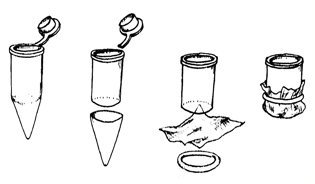
Figure 23. Holders for embryos.
Plastic microwells (0.5 ml volume) can be used to hold solutions. Add fresh solutions by pipetting them on top of the tissue. When transferring holders from one well to the next, drain mesh bottom briefly on Kimwipes®. Small plastic Petri dishes or Stender dishes can also be used, in which case several holders can be processed at the same time.
The stained embryos can be transferred to glass spot dishes for dehydration, removing the yolk if desired while transferring through alcohols. It is also possible to leave the tissue in the BEEM capsule holders while dehydrating.
Some flexibility in the staining schedule is possible. Thorough washing seems to be the most important factor in reducing background and should not be minimized.
Staining of 30 hpf fish or older is improved by adding Triton X-100 to a final concentration of 0.1% to 0.5% to all solutions.